Canary Center at Stanford Resource Facilities
Additive Manufacturing Prototyping Facility (AMPF)
The Additive Manufacturing and Printing Facility (AMPF) at Stanford aims to establish leadership in the future of manufacturing by providing a space for faculty, students, and entrepreneurs to turn their ideas into market-ready products. This advanced 3D printing facility will enable digital design, prototyping, and small-scale production as an integrated innovation hub and shared use facility that will be a key pillar for innovation and commercialization for investigators spanning the Stanford campus and Silicon Valley.
Cell / Molecular Biology (CMB) Resource Facility
The Cell and Molecular Biology Shared Resource Facility (CMB Resource) at the Canary Center facilitates the development of tools for early diagnosis of cancers. The CMB Resource is well equipped for developing molecular imaging probes. Specifically, this resource develops and characterizes antibody and ligand-based probes for targeted molecular imaging thus aiding faculty, scientists and students at the Center who focus on developing highly sensitive multifunctional optical, PET and MRI probes for imaging cancers by targeting cancer-specific cellular targets.
The CMB Resource houses a variety of instruments needed for performing highly advanced molecular biology experiments. The instruments used by the resource include, for example: a highly sensitive fluorescent microscope with a time-lapsed imaging functionality, a real-time PCR machine, a microplate reader with fluorescence and bioluminescence capabilities, a fluorescent cell sorter, an IVIS-Lumina –highly sensitive cooled charge coupled device camera for imaging bioluminescence and fluorescence of different wavelengths, and an automated peptide synthesizer. The CMB Resource is also equipped with a tissue culture facility that includes BSL-2+ capabilities.
The goal of the CMB Resource mimics that of the Center in that it aims to promote the development and advancement of methods for cancer early detection. The Cell and Molecular Biology Shared Resource Facility works closely with Center Members of the other Shared Resources to forward the Center’s research efforts.
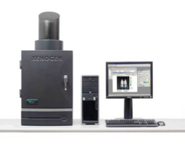
IVIS-Lumino-high sensitive cooled charge coupled device camera for imaging bioluminescence and fluorescence intact cells and non-invasive imaging in live animals.
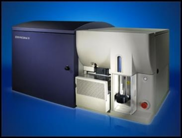
The state of the art BD FACS Aria II cell sorter which enables high-throughput screening of large biomolecular libraries and selection of high affinity binders from yeast and mammalian cells
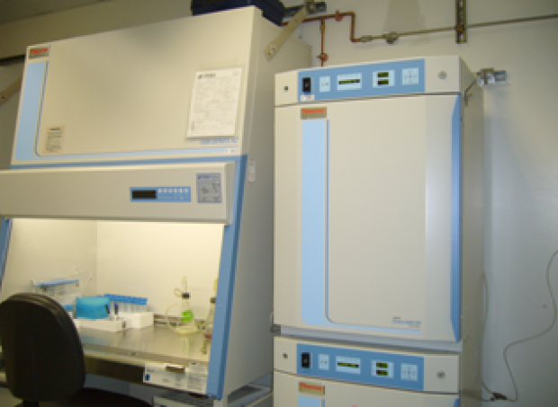
State of the art-cell culture facility.
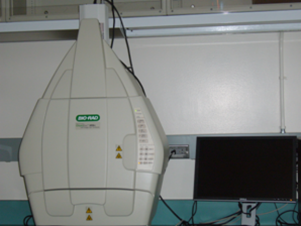
UV-Geldoc system with chemiluminescence imaging facility.

Automated peptide synthesis allows rapid validation of new probes engineered to bind cancer targets with high affinity and specificity.
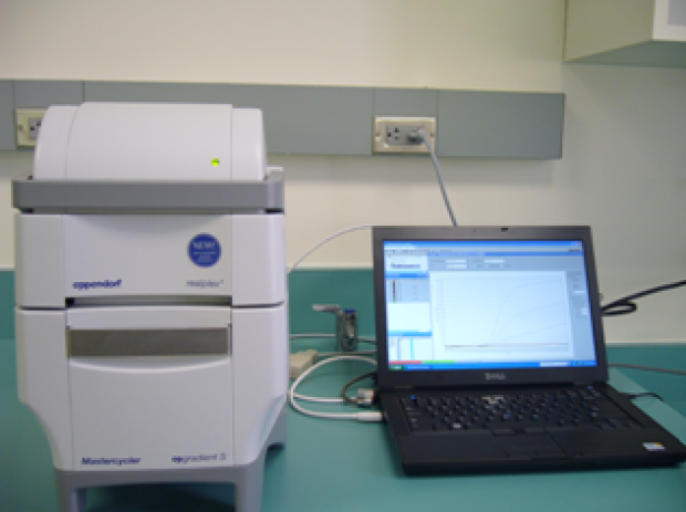
Real time PCR-system for the rapid quantitation of gene expression and microRNA from cells and tissues.
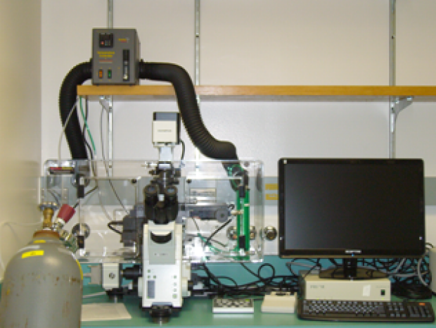
High sensitive fluorescent microscope with weather controlled FRET and time lapsed imaging facility.
Center for STEMM Mentorship
The mission of the Stanford Center for STEMM Mentorship is to cultivate the well-being, scientific growth, and personal and professional development of members of STEMM research teams—students, postdocs, staff, and faculty, all together—by advancing whole person-centric, team-oriented mentorship rooted in principles that drive successful academic, industrial, entrepreneurial, and athletic teams.
Preclinical Imaging Core Facility

Edwin Chang
Director, Preclinical Imaging Core Facility
E-mail »
The mission of the preclinical imaging facility is to develop infrastructure, expertise and tools to support customized in vivo imaging as well as image and data analysis for direct use by the basic, clinical and translational researchers located at Porter Drive and/or nearby Stanford Labs. As a shared resource facility within the Canary Center at Stanford for Cancer Early Detection, the core also supports Canary Center Members, Associate Members, and Canary Foundation Science Teams
Part of the Stanford Center for Innovation in In-vivo Imaging (SCI3)
As an expansion to the Stanford Center for Innovation in In-vivo Imaging (SCI3), the Preclinical Imaging Core at Porter Drive was established to support the Canary Center at Stanford for Cancer Early Detection. Its mission is to provide access to state-of-the-art and first-of-its-kind preclinical imaging instruments to facilitate the translation of research from in vitro tests to small animal investigations and clinical practice. This enables evaluation and advancement of novel imaging technologies and biological concepts in living murine models. Researchers are trained in the use of each of the imaging modalities available, and several facilities are located on the Stanford campus (at The James H. Clark Center, Lorry I. Lokey Stem Cell Research Building, Comparative Medicine Pavilion, and Shriram Center for Bioengineering & Chemical Engineering) to enable ease of access for scientists and their research animals.
The Preclinical Imaging Core provides access to a spectrum of imaging modalities, including instruments routinely found in hospitals but optimized for small animal work (such as ultrasound, MRI, CT, and PET), instruments developed specifically for small animal work (such as optical imaging), as well as new equipment that has just been developed (such as photoacoustic imaging). All instruments are designed to image living subjects and allow for repeated imaging, which reduces the number of animals that researchers need to use. The flexibility and rapid analyses of such animal models greatly accelerate the development of molecular imaging strategies. The facility also houses a dedicated surgical procedure room, a histology slide scanner that converts glass slides into digital slides using both brightfield and fluorescence, and several advanced image analysis workstations.
The Preclinical Imaging Core operates as a Stanford School of Medicine Service Center and is supported by the Canary Center at Stanford, the Stanford Cancer Institute, as well as user fees. It is operated by the Departments of Radiology and Pediatrics. Edwin Chang is the director of the Preclinical Imaging Core at Porter Drive and Dr. Heike Daldrup-Link oversees the entire Stanford Center for Innovation in In-vivo Imaging (SCI3).
Access to the imaging facility is restricted to the authorized users only. Contact Edwin Chang for more information about how to obtain access to the facility and get hands on training or other services. As a service center the imaging facility will collect fees from users based on the Stanford shared resource model. Faculty who are interested in joining the facility should contact Frezghi Habte for more information.
The imaging facility currently houses nine instruments in category of six different modalities. Our goal is to provide up-to-date instrumentation for preclinical imaging and we will continue acquire new instruments when funds are available. Below is a brief description of each of the available instruments.
Positron Emission Tomography (PET) is an imaging system that images high-energy γ-rays emitted from within the subject. The source of imaging for PET comes from the injected positron emitting biological molecules or radiotracer such as 18F-FDG (fludeoxyglucose) into the subject. As the radioisotope decay, it emits positrons, which annihilates with electrons found naturally in the body. This produces 2 γ-rays at ~180° apart and detected by sensors on opposite ends of the PET machine. This allows individual emission events to be localized within the body, and the data set is reconstructed to produce images of the distribution of the injected radiotracer within the subject. Non-invasive, in vivo functional imaging with microPET allows both serial and longitudinal studies to be conducted in the same animal. We have two different MicroPET systems at the imaging facility located at Porter.
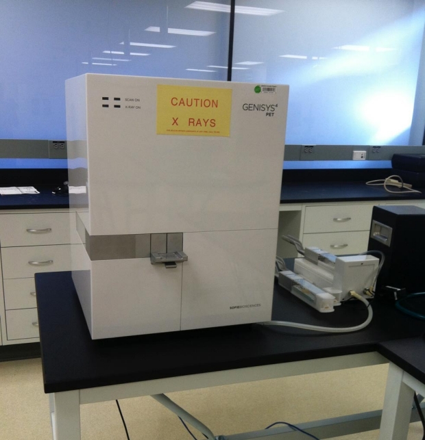
G4 PET/X-Ray System
A tablebench MicroPET system that allows a quick scan for mice and small rats with fully–integrated docking stations/imaging chambers and user-friendly acquisition/reconstruction software platform. G4 PET/X–Ray is built from four panel detectors that surround the animal (mouse full body and rat head/trunk) with a novel system model and iterative reconstruction. X-ray is included to provide anatomical reference for co-registration of the PET images with built in digital image of the mouse anatomy.
Key features/specs G4 PET/X-RAY system are:
- Ultra high sensitivity 14%
- Spatial resolution 1.4 mm
- Crystal dimension - 1.4x1.5x10 mm^3
- Field of View, Transaxial – 100 mm, Axial – 127 mm
- Animal monitoring system
- Heated Custom imaging chamber
- Controllably anesthesia system
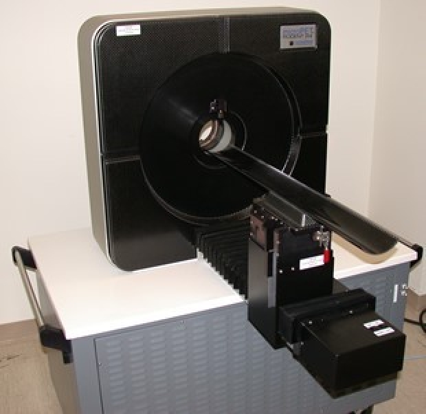
Siemens MicroPET R4
One of the first commercial systems with a 19 cm (transaxial) by 7.6 cm (axial) field of view. The large transaxial field of view allows multiple animals to be scanned simultaneously. With multi-mice holder developed in-house, we currently are routinely imaging four animals at the same time increasing the through of the system. The system has an absolute sensitivity of 4% with a spatial resolution of ranging from 1.3 at the center to 2 mm at the outer edge of the field of view. List-mode data acquisition allows acquired data to be processed in a static or dynamic data set to provide quantitative snapshots or summed sequences of radioactivity distribution in live animals. An interchangeable bed allows successive PET and other modality such as CT or MRI scans of the small animals to provide co-registered structural and metabolic images similar to scans performed with dual modality MicroPET/CT or MicroPET/MRI machines
Historically, optical imaging as the first imaging device uses visible light as source of imaging but with very limited tissue penetration. The most common optical imaging modalities used for in vivo small animal imaging are the fluorescence and bioluminescence imaging systems. Fluorescence imaging gets its source for imaging from fluorochromes injected inside the subject that are excited by an external light source and emitting light of a different wavelength in response. Traditional fluorochromes include GFP, RFP, and their many other mutants. Recently, near-infrared dyes and infrared fluorescent proteins (700 nm-800 nm) are used due to low autofluorescence and deeper tissue penetration at these wavelengths. Bioluminescence imaging, on the other hand, is based on light generated by chemiluminescent enzymatic reactions such as luciferase. In both fluorescence and bioluminescence, the light signals are captured by Charged Coupled Device (CCD) cameras cooled up to -95 °F to be extremely light sensitive. Three optical imaging systems are currently available at the porter imaging facility.
Spectral Instruments Imaging (SI Imaging), Lago
is a second-generation optical imaging platform for general purpose optical imaging. Lago uses a back-illuminated CCD air-cooled to -90°C camera. The system can perform both bioluminescence and fluorescence imaging. The system features quantitative and analytical software to capture and analyze images.
Key features:
Pixel dimensions |
2048x2048 |
Pixel size |
13.5×13.5um |
CCD size |
27.6x27.6um |
Quantum efficiency |
>85% from 500-650nm, >30% from 400-850nm |
Dark current |
<0.00009 e-/pixel/s |
Binning |
1x1, 2x2, 4x4, 8x8, 16x16 |
Lens |
50mm, min aperture f/1.2, max f/16 |
Imaging Field of View |
25x25cm max, 6x6cm min |
Fluorescence excitation filters |
14 |
Fluorescence emission filter slots |
20
|
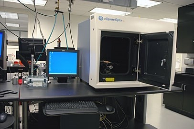
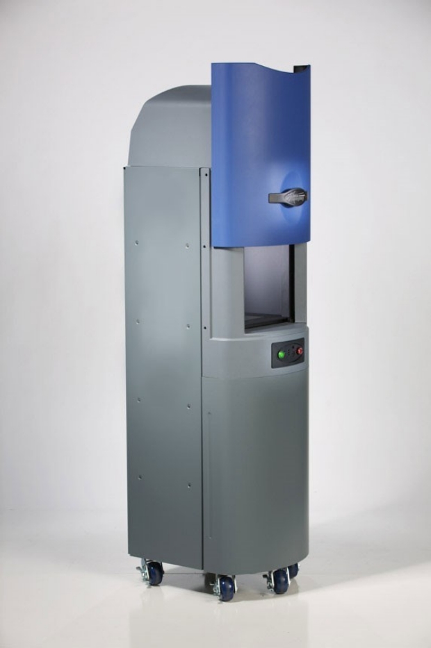
Art Optix
A fluorescent molecular imaging system based on time-domain technology. It has the capacity to image red fluorescent proteins and Q-dots. The system can also retrieve fluorescence lifetime and features a CT-fusion software application.
Ultrasound imaging systems works through the generation of sound waves from transducers into the subject. As the sound waves propagate through tissue, they are reflected back and picked up by the transducer, and can then be translated into 2D and 3D images. Ultrasound systems for small animals are specifically developed with higher frequencies ranging from 15 MHz to 80 MHz compared to clinical ultrasound system with frequencies range from 3-15 MHz. In addition, contrast agents in the form of microbubbles, which have different acoustic properties from that of tissues can be introduced into animal systems to highlight structure such as vasculature or be targeted towards specific receptors.
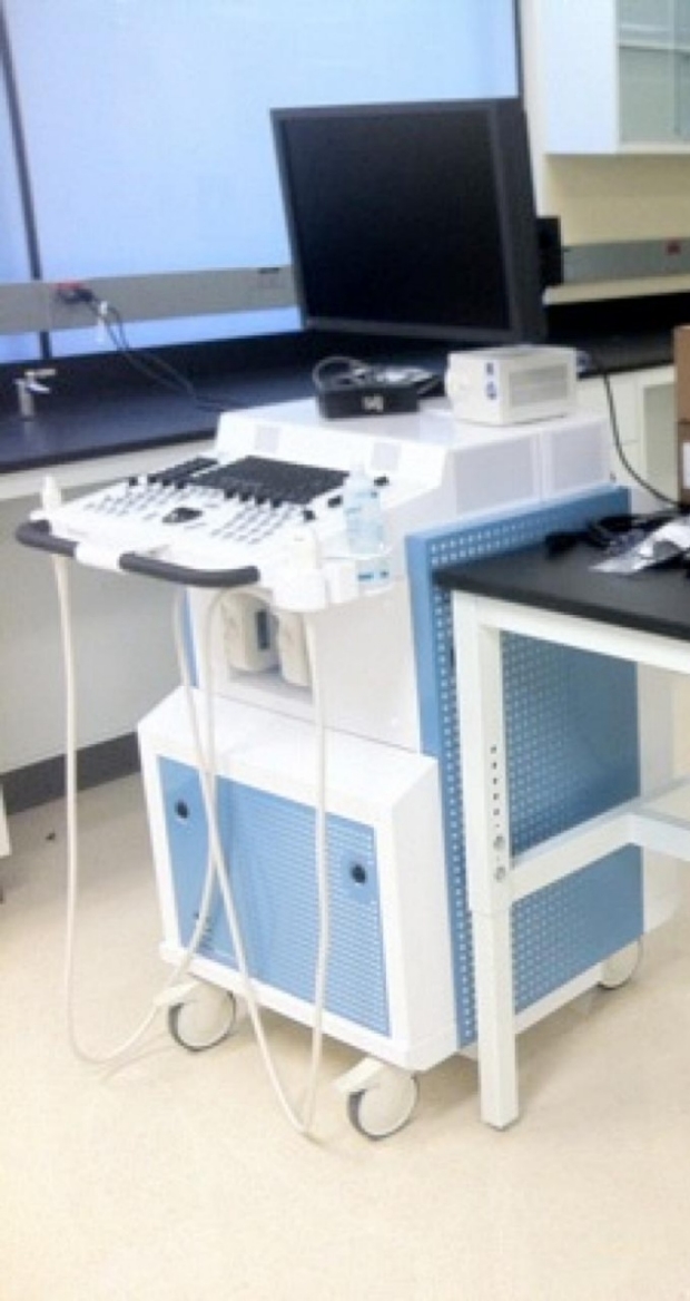
Vevo 2100 System
The next generation ultrasound system developed to expand the functionality, flexibility and image quality of the older Vevo 770 system. It operates at higher frequencies using new solid-state array transducers. The new MicroScan™ transducers provide increased frame rates, higher contrast and resolution, and a wider field of view. The system is easy to use, non-invasive and fast, providing extremely high throughput when needed.
Key features
- Superior resolution, 30 microns
- Uniform image quality through entire FOV
- Color and Power Doppler Modes for blood flow quantification & anatomical identification
- M-Mode single line acquisition allowing high-temporal resolution for LV functional analysis
- 3D-Mode Imaging & Volume Analysis
- Nonlinear Contrast Imaging
- Advanced measurements & quantification
Photoacoustic imaging is a technique that combines optical illumination, optical absorption, and ultrasound detection to map the location of absorbers within light scattering media with high sensitivity and spatial resolution. A short pulse of electromagnetic radiation induces a milli-Kelvin rise in temperature within the absorber resulting in a thermal expansion. The rapid heating and cooling results in an acoustic wave that is emitted from the absorber that may be detected at the tissue surface by acoustic receivers. The depth of the absorber may be determined by the time of arrival of the thermo-acoustic signal at the tissue surface. A typical photoacoustic imaging system employs a laser source that generates pulses of light <10 nsec in duration with pulse energy in the 1-30 mJ range. Acoustic receivers are typically wideband and have center frequencies in the 1-20 MHz range.
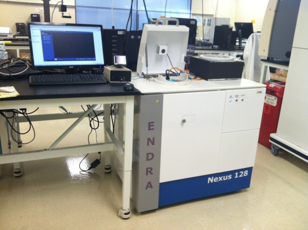
Photo-Acoustic Enhanced Ultrasound, Nexus 128
This system can perform 3D photoacoustic computed tomographic (CT) imaging. Tomographic images of optical absorption are generated using endogenous and/or exogenous contrast. Photoacoustic CT is suitable for contrast enhanced and molecular imaging applications through the use of near infra-red absorbing dyes or fluorescent probes.
X-Ray Computed Tomography (CT) uses X-ray imaging to produce cross sectional and volumetric images of the subject. X-rays are projected through the body onto an array of detectors that measure the X-ray attenuation along the X-ray source and detector. The source and detector are then rotated around the subject to make multiple views at different angles of rotation. Using computer reconstruction algorithms, an estimate of absolute density at each point (volume element, “voxel”) in the body is computed. Thus, the CT image is a computed image of the measured X-ray attenuation from all projects. MicroCT is a miniaturized version of the conventional CT imaging system. The MicroCT currently available at Porter preclinical imaging facility is a Viva CT 40 scanner from Scanco Medical.
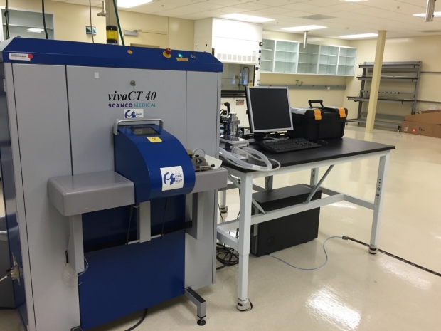
Scanco Medical VivaCT 40
A high-speed in vivo micro CT scanner ideal for resolving dense tissues, including bone and contrast enhanced structures such as vasculature. It is equipped with a microfocus X-ray source capable of 5 μm spot size and 50-70 kVp, 8W (160 μA) and can achieve up to10 μm nominal resolution. It can cover a maximum scan length of 145 mm and has axial field-of-view up to 20-38 mm. Samples can be up to 80 mm in diameter and up to 200 mm in length. The system includes software capable of data acquisition, online or offline reconstruction, 2D & 3D analysis and volume reconstruction, 3D visualization and animation, and also data archiving.
Coming soon
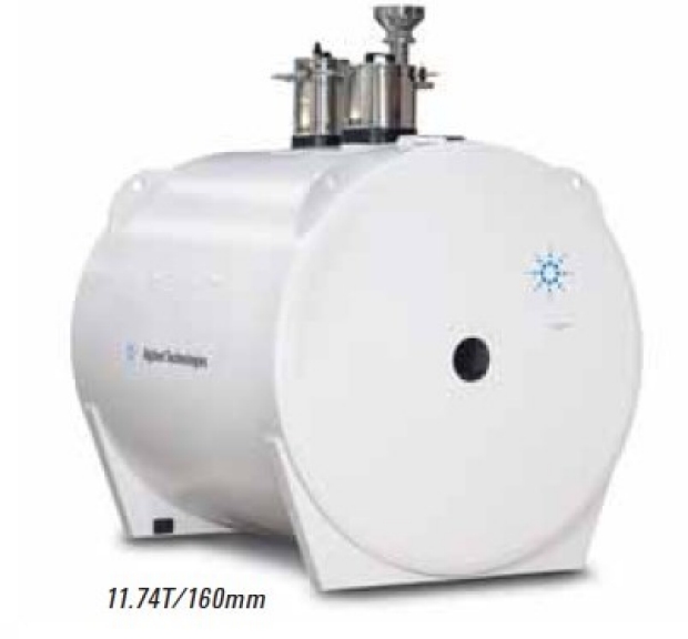
The imaging facility at Porter currently is currently in the process of installing high field small animal MRI system, Agilent 11.7T MRI.
MRI exploits the nuclear magnetic alignments of different atoms inside a magnetic field to generate images. MRI machines consist of large magnets that generate magnetic fields around the target. A radiofrequency (RF) coils inside the MRI generates radio pulse of particular frequency that cause paramagnetic atoms such as hydrogen, gadolinium, and manganese to align in a magnetic dipole along the magnetic fields. When the RF pulse is temporarily ceased the atoms return to original aligned state (a process called “relaxation”), emitting a detectable radiofrequency signal that is captured by the MRI system. With this data (referred to as T1 and T2 relaxation), a computer will generate an image of the subject based on the resonance characteristics of different tissue types. Differences of T1 and T2 are inherent properties of tissue and they vary among tissues. Thus, MRI provides detailed functional information about tissue and can be valuable for in vivo imaging providing high tissue contract. With high magnetic field and flux radiofrequency waves, MRI can also give high spatial resolution. MRI can be used in a wide variety of applications, including anatomical, functional, and molecular imaging. Furthermore, since MRI’s mechanism is based on a magnetic field, it is much safer compared to radiation based imaging modalities such as CT and PET.
Computational Resources
The image facility also provides computational resources for data analysis. Three workstations are currently available for general computational and data analysis use with the following image/data analysis software packages.
- Workstation 7 (Mac): OsiriX, ImageJ and Matalb
- Workstation 8 (PC): IRW(Inveon), Living Image, GE MicroView, Amide, Amira, ImageJ and Matlab.
- Workstation 9 (PC): IRW (Inveion), vivoQuant, Living Image, GE MicroView, Amide, ImageJ and Matlab
Other Equipment
The imaging facility is also equipped with other supporting equipment and tools for imaging. Some of these include:
- Four surgical stations with anesthesia system for performing basic surgical procedures
- Well counter, Dose Calibrator and Gamma Counter for radio isotope counting and measurements
- Surgical microscopy (Leica).
General policy / Pricing
General policy for instrument use
- Access. Access to the imaging facility and all equipment must be approved by the facility Director. Approved users will have their ID cards activated to access the imaging facility and be included on instrument booking system.
- Training. All users must receive proper training before being permitted access to the equipment needed. Training is provided by the director and can be requested by contacting Edwin Chang at echangcv@stanford.edu
- Scheduling. Advance reservation using our online scheduling calendar is required. Cancellation at least 4 hours prior to the experiment is required. Users failing to do this will be charged (at the hourly rate) for the booked time. Contact the facility staff if you need to cancel after the 4-hour deadline.
- Radioactivity and Safety. Users are responsible for their compliance with the Stanford Safety Policy.
- Billing. Users will be required to provide billing information prior to requesting instrument time. Users with multiple billing accounts should specify the account number to be charged upon sign-up..
- Cleaning Up. Users are responsible for keeping the equipment and work areas safe and very clean.
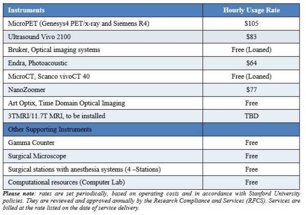
Pricing
As a Service Center, the imaging facility will operate on a fee-for-service basis. Usage fees are calculated at per usage hour base and include reconstruction of datasets in standard format. Instrument use for post-processing will not be charged, provided that it is done when the instrument is free without interfering with any other scheduled studies. The service center does not provide radioisotopes and contrast imaging agents and these costs are not included in the instrument usage fees.
Table below lists the currently available instruments/services and their hourly rates, including the instruments available free of charge to support users at the imaging facility
Frezghi Habte, PhD
Director, Preclinical Imaging Core Facility
E-mail »
Please email Frezghi Habte, PhD (fhabte@stanford.edu) to initiate an instrument training request from the Preclinical Imaging Core. Please include your name, contact information and a short description of your request. Dr. Habte will respond within 48 hours.
Proteomics Resource Facility
For more information regarding the Proteomics Resource Facility, Please contact Abel Bermudez
The Proteomics Shared Resource Facility (Proteomics Resource) associated with the Canary Center is a shared resource that supports Canary Center Members, Associate Members, and Canary Foundation Science Teams. The facility supports the mission of the Center to foster research leading to the development of blood tests and molecular imaging approaches to detect and localize early cancers.
The facility houses mass spectrometry (MS), liquid chromatography (LC), and sample preparation systems employed primarily for biomarker discovery and verification studies. Stanford researchers with interests in cancer early detection seeking access to the resources available in the facility should do so in collaboration with Canary Center Members. At present, the Proteomics Resource supports four mass spectrometers including: (1) Thermo LTQ Orbitrap Elite; (2) Thermo LTQ Orbitrap Velos; (3) Agilent 6490 triple quadrupole; and (4) AB SCIEX 5800 TOF/TOF (MALDI) systems. Other instruments available in the shared resource include an Agilent AssayMAP Bravo liquid-handing robotic system, FortéBio Octet 384 system for biomolecular interaction analysis, as well as several systems for peptide and protein fractionation and sample preparation.
In general, biomarker research follows a continuum that begins with discovery and proceeds through validation to the eventual implementation of biomarkers in a clinical setting. Biomarker discovery requires high confidence identification of biomarker candidates with simultaneous quantitation information to indicate which proteins are changing to a statistically relevant degree in response to disease. Biomarker candidates identified in discovery need to be validated using larger sample sets covering a broad section of patient cohorts. To avoid a potential bottleneck associated with taking a large number of candidates to validation, a verification step is employed to screen potential biomarkers to ensure that only the highest quality leads from the discovery phase are taken into the costly validation stage. The verification stage requires a high throughput workflow with a minimum of sample preparation that provides both high specificity and sensitivity. Additionally, the verification stage can confirm that a particular methodology is suitable to be used in the validation phase.
Biomarker Discovery Workflows
One of the biggest challenges in biomarker discovery is the difficulty of identifying medium to low abundance proteins in complex biological samples. For example, human plasma has over 1 X 10⁶ different protein molecules with an estimated dynamic range approaching 1010. Of these, the 22 most abundant proteins make up 99% of the protein content of plasma, and the abundance of a given biomarker will fluctuate within a sample population. A robust biomarker discovery workflow must be capable of uncovering a panel of biomarkers in samples such as human plasma and cancer cell line lysates, with unambiguous identification and quantitative characterization. To address this challenge, quantitative proteomics workflows involving SILAC (Stable Isotope Labeling of Amino Acids in Culture) are employed. In the core facility, chiefly the Thermo Orbitrap Elite and Thermo LTQ Orbitrap Velos LC/MS systems are used for biomarker discovery.
Biomarker Verification Workflows
Because of normal clinical or biological variability, candidate biomarkers identified in the discovery stage need to be validated across a large number of samples. The challenge is to develop a fast, targeted analysis method capable of analyzing as many identified candidates as possible in minimally hundreds and potentially even thousands of samples. A biomarker candidate verification phase eliminates this bottleneck by ensuring that only the most promising putative biomarkers found in discovery go on to the validation stage. To address this challenge, targeted proteomics workflows involving MRM (Multiple Reaction Monitoring) and SISCAPA (Stable Isotope Standard with Capture by Anti-Peptide Antibodies) are employed on the Agilent 6490 iFunnel triple quadrupole LC/MS system.
Workflow Tutorials
Targeted LC/MS Methods
MALDI Applications
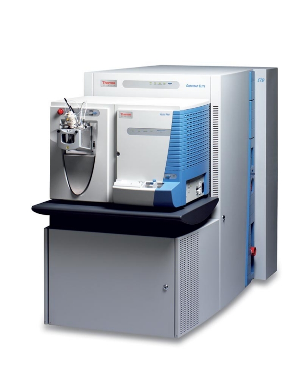
Thermo Orbitrap Elite with Electron Transfer Dissociation (ETD) and Thermo NanoMate 3000 UHPLC
The Thermo Scientific Orbitrap Elite hybrid ion trap-Orbitrap mass spectrometer provides the highest resolving power of all Orbitrap-based instruments of up to 240,000 FWHM at a scan rate of 4 Hz, to achieve ultimate detection limits and selectivity in the full-scan MS for analysis of complex samples and providing accurate quantitation results. MS/MS in the ion trap allows probing the least-abundant components in a complex mixture. Fast, high-quality MS and MS/MS scans allow maximal identification of peptides in a complex matrix. Additionally, multiple fragmentation techniques, including collision-induced dissociation (CID), higher-energy collisional dissociation (HCD) and electron transfer dissociation (ETD), provide complementary fragment information for maximal sequence coverage.
Specifications*
Mass Range Dynamic Range MS Scan Power Mass Accuracy Resolution 50 to 2000 m/z; 200 to 4000 m/z > 5000 within a single scan MSn for n = 1 through 10 < 3ppm RMS with external calibration < 1ppm RMS using internal calibration 60,000 at m/z 400 at a scan rate of 4Hz Minimum resolution 15,000 at m/z 400 Maximum resolution > 240,000 at m/z 400 *Thermo Scientific Orbitrap Elite Product Specifications #PS30229 (05/11)
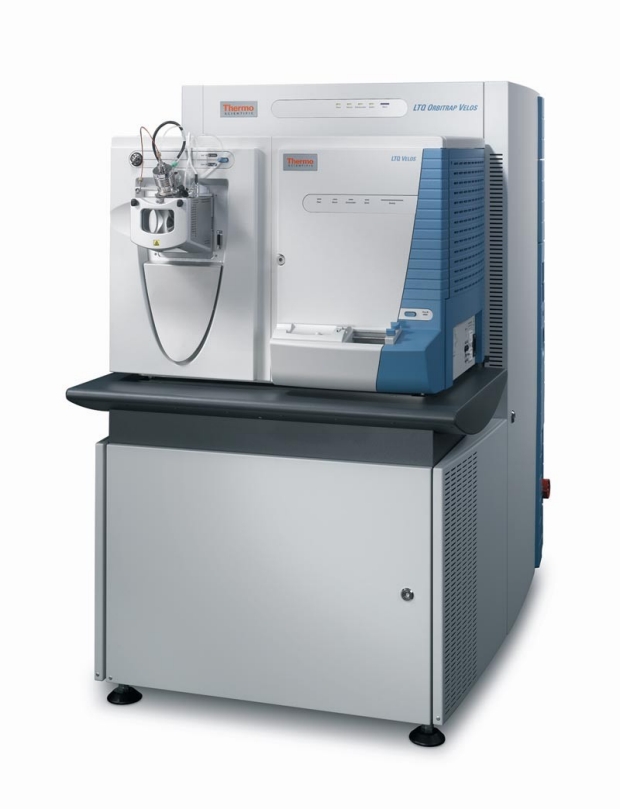
Thermo LTQ Orbitrap Velos with Eksigent NanoLC-2D HPLC
The Thermo Scientific LTQ Orbitrap Velos mass spectrometer combines the mass accuracy and ultra-high resolution of the Orbitrap mass analyzer with resolving power of up to 100,000 FWHM at a scan rate of 1 Hz, with the increased sensitivity and improved cycle time of the LTQ Velos dual-pressure linear ion trap, to achieve excellent detection limits and selectivity in the full-scan MS for analysis of complex samples and providing accurate quantitation results. MS/MS in the ion trap allows probing the least-abundant components in a complex mixture and high-quality MS and MS/MS scans allow maximal identification of peptides in a complex matrix.
Specifications*
Mass Range Dynamic Range MS Scan Power Mass Accuracy Resolution 50 to 2000 m/z; 200 to 4000 m/z > 5000 within a single scan MSn for n = 1 through 10 < 3ppm RMS with external calibration < 1ppm RMS using internal calibration 60,000 at m/z 400 at a scan rate of 1Hz Minimum resolution 7500 at m/z 400 Maximum resolution > 100,000 at m/z 400 *Thermo Scientific LTQ Orbitrap Velos Product Specifications #PS30185 (05/09)
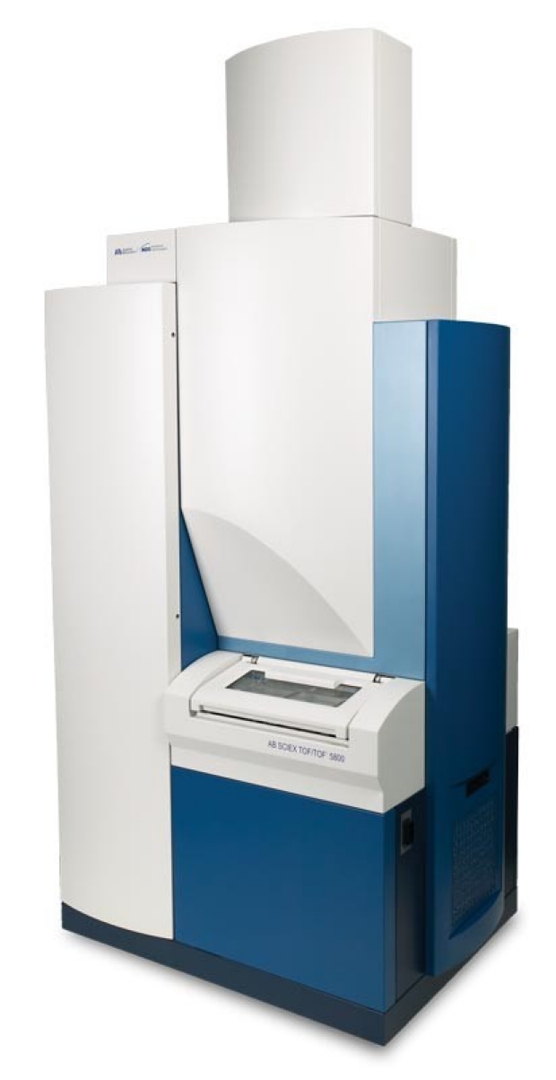
AB SCIEX 5800 TOF/TOF System
The AB SCIEX 5800 TOF/TOF system provides a confident path to protein identification and relative quantitation for occasional users who wish to acquire proteomics data to complement other approaches (genomics, metabolomics, etc.). The system's speed and sensitivity make it ideal for analysis of intact proteins purified by electrophoresis and column chromatography as well as relative quantitation of peptides in a digest using iTRAQ isobaric tagging. Electrospray ionization (ESI) and MALDI have been shown to provide complementary information for peptide identification. MALDI TOF/TOF data can be used to complement ESI data for a more in-depth identification and quantitation of proteins. Furthermore, the LC-MALDI workflow separates the time constraints of liquid chromatography (LC) from the mass spectrometry. With your chromatographic run "stored" on a MALDI plate, enabling subsequent re-analysis, and the 5800 TOF/TOF system automatically spends the time required to get the maximum information from any time point in the chromatographic separation.
Specifications*
Laser Repetition Rate Mass Range Mass Accuracy Resolution Resolution (broad mass range) MS, 200 and 400 Hz; MS/MS, 200, 400 and 1000 Hz 100 to 250,000 m/z (up to 1.2 MDa with COVALX HM2 Detector) < 10 ppm RMS with external calibration < 2.5 ppm RMS using internal calibration 30,000 for single peak in mass range 1200 to 3700 m/z > 16,000 for Angiotensin 1 > 18,000 for Glu1-Fib B > 25,000 for ACTH 1-17 and ACTH 18-39 > 18,000 for ACTH 7-38 *Provided by AB SCIEX (unpublished)
COVALX HM2 High Mass Detector
The AB SCIEX 5800 TOF/TOF system has been fitted with a COVALX HM2 High-Mass Detector that enables detection of macromolecular ions up to 1.2 megadaltons (MDa) with nM sensitivity. The HM2 high-mass detector retrofit does not alter any of the features of the MALDI instrument, and both the standard linear and reflectron detectors remain available. The high-mass detector supports a number of important applications including
- intact protein complex analysis,
- antibody characterization, and
- therapeutic protein aggregation analysis.
AB SCIEX Tempo LC-MALDI Spotting System
The Tempo LC-MALDI Spotting System provides a high precision plate spotting technology. It provides contact-free electrostatic deposition of droplets and includes a three-channel micro-LC pump for the LC gradient and post-column addition of MALDI matrix. For greater throughput and automation, the system accommodates multiple plates and multiple LC runs per plate. An integrated autosampler and low-dispersion UV detector are also included. LC-MALDI frees MS analysis from the time constraints of LC to enable more in-depth interrogation of samples, which can be of particular advantage in applications involving intact protein separations as well as analysis of tryptic digests. The Tempo LC-MALDI system dispenses droplets on plates by applying a high voltage electrostatic pulse on the sample needle at controlled intervals. This non-contact deposition technique virtually eliminates any occurrence of missed or misaligned spots and routinely delivers highly reproducible results and high-quality MS data. Spot coordinates are transferred directly to the AB SCIEX 5800 TOF/TOF system to facilitate mass spectrometry.
SimulTOF 200 Combo MALDI-TOF
The SimulTOF 200 Combo mass spectrometer is the highest performance bench top reflector system available. It is suited for intact protein, peptide and small molecule analysis. It is particularly well suited for imaging mass spectrometry (IMS). A high performance hybrid linear detector provides excellent mass range (100 Da to 500 kDa), speed (5 kHz, up to 100 spectra/sec) and dynamic range. A second high performance microchannel plate detector is used in the reflector mode. The SimulTOF 200 Combo was designed for ease-of-use through intuitive software, and low maintenance by exploiting new concepts in instrument design. The plate editor allows the user to specify the geometry of any plate the system might support including conventional plates as well as microscope slides for IMS.
Specifications*
20 kV source and novel high speed, high mass detector provides very high sensitivity, resolving power, and accuracy over a broad mass range. High laser rate (5 kHz) and high acquisition rate (up to 100 spectra/sec) makes tissue imaging practical. Resolving power in Linear Mode: >6000 for ACTH @ 2465 Da; Resolving power in Reflector Mode: >20,000 for Neurotensin @ 1672.92 Da and >10,000 for full range of from 1000 Da to 1 kDa. Mass range: 100 Da to 500 kDa. Sensitivity, Linear Mode: S/N>10 for ACTH (2465 Da) @ 1 attomole/mL and S/N>10 for IgG (150,000 Da) @ 1 fmole/mL. Sensitivity, Reflector Mode: S/N>10 for ACTH (2465 Da) @ 1 fmole/mL. Mass Accuracy, Reflector Mode: <5ppm across the sample calibrating on a single spot.
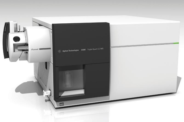
Agilent Technologies 6490 Triple Quadrupole LC/MS
Incorporating breakthrough iFunnel technology, the Agilent 6490 Triple Quadrupole LC/MS takes detection limits lower than ever. Users can achieve zeptomole level sensitivity and an unprecedented six orders of linearity for the most demanding quantitative analyses. Proprietary iFunnel technology dramatically increases ion sampling and transmission efficiency, enabling the lowest limits of detection and quantitation. Triggered MRM combines fast and sensitive MRM quantitation with the generation of a product ion spectrum for library searching, compound screening and confirmation. MassHunter Optimizer software automatically optimizes MRM transitions and fragmentor and collision energies for both small molecules and peptides. Ultra fast MRM acquisition speeds and ion polarity switching, an innovative collision cell eliminates cross-talk and enables 1ms MRM dwell times, together with fast polarity switching, ensures compatibility with UHPLC analyses. Dynamic MRM simplifies method development and allows thousands of compounds to be quantified in a single method with optimal sensitivity and reproducibility. Comprehensive and easy-to-use MassHunter Workstation software dramatically enhances productivity from method optimization, data acquisition to data processing and reporting.
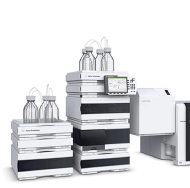
Agilent 1260 Infinity HPLC-Chip Cube MS Interface
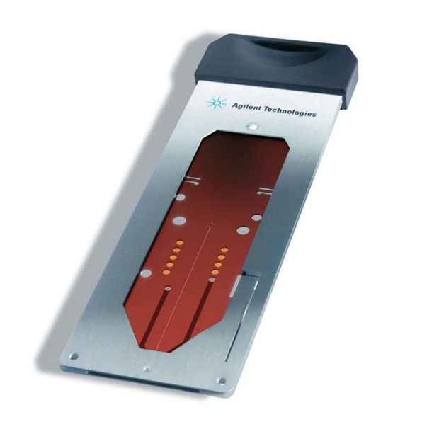
The Agilent 6490 Triple Quadrupole LC/MS may be configured with the Agilent 1260 Infinity HPLC-Chip Cube MS Interface, a microfluidic chip-based technology specifically designed for nanoelectrospray LC/MS. Based on Agilents laser etched polyimide HPLC-Chip and the HPLC-Chip Cube MS interface, the Agilent HPLC-Chip/MS system affords nanoelectrospray MS sensitivity, robustness, reliability, and ease of use. Reusable HPLC-Chips seamlessly integrate sample enrichment and separation nanocolumns, tubing, connections, and spray needle of a traditional nanoelectrospray LC/MS system into a biocompatible polymer chip. This results in narrower, better-defined peaks due to elimination of post-column dead volumes. HPLC-Chip Cube provides easy to use operation with automatic chip loading, solvent and sample delivery to the chip, high pressure switching of flows and automated chip positioning in the MS source. In the Proteomics Core, the HPLC-Chip Cube MS interface is used with Agilent 1260 Infinity series nanoflow and capillary pumps. When combined with the Agilent 6490 Triple Quadrupole MS, the HPLC-Chip for LC/MS offers even greater overall reliability and ease-of-use for many LC applications. HPLC-Chips with novel surface chemistries are currently available for protein ID, biomarker discovery and verification, phosphopeptide analysis, glycan analysis, and intact protein analysis.
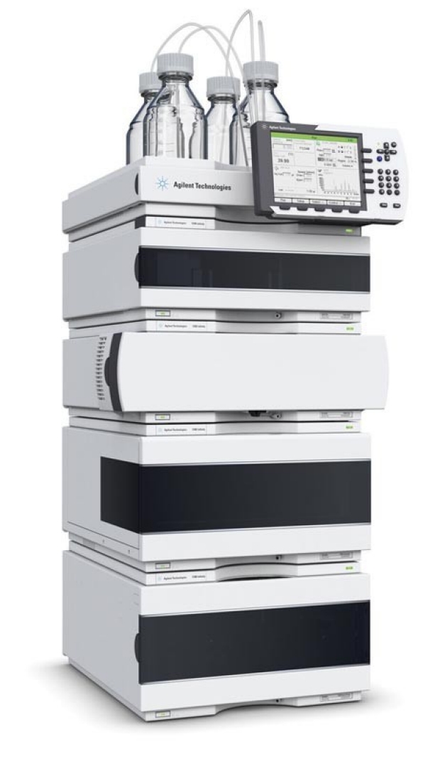
Agilent 1290 Infinity Series UHPLC
The Agilent 6490 Triple Quadrupole LC/MS may be configured with the Agilent 1290 Infinity Series UHPLC (ultrahigh performance liquid chromatograph). The Agilent 1290 is designed to provide highest speed, resolution and sensitivity. The infinite power range that combines ultra high pressure up to 1200 bar and high flow rates up to 5 mL/min for maximum chromatographic performance, compatibility, and flexibility allows enables the user to deploy any particle type, any column dimensions, or any mobile and stationary phases. Innovative technology components offer excellent performance for both UHPLC and HPLC applications. The 1290 Infinity Series UHPLC is the first system that provides the foundation for method transfer to or from any Agilent or non-Agilent UHPLC or HPLC system. Lab Advisor software with intuitive diagnostic and monitor capabilities and alert functions notifies users of problems.
Agilent 1290 Infinity Series UHPLC with Variable Wavelength Detector
The Agilent 1290 Infinity LC is an UHPLC (Ultra-High Performance Liquid Chromatograph). The 1290 LC is designed to provide highest speed, resolution and sensitivity. The infinite power range that combines ultra high pressure up to 1200 bar and high flow rates up to 5 mL/min for maximum chromatographic performance, compatibility, and flexibility allows enables the user to deploy any particle type, any column dimensions, or any mobile and stationary phases. Innovative technology components offer excellent performance for both UHPLC and HPLC applications. Lab Advisor software with intuitive diagnostic and monitor capabilities and alert functions notifies users of problems. The Agilent OpenLAB Chromatography Data System (CDS) workstation and the Agilent 1260 Infinity Variable Wavelength Detector (VWD) enable the 1290 LC to operate as stand-alone analytical UHPLC. The VWD is highly sensitive for HPLC and RRLC analyses. It uses time-programmable wavelength switching so the user can optimize the detection sensitivity and selectivity for specific applications. The latest electronics and refined optics have lowered detector noise levels to below 2.5 μAU, and the lowest baseline drift (< 1 x 10⁻⁴ AU/h) means users can precisely quantify trace level compounds in samples. The 1290 Infinity Autosampler provides fast sample injection with highest precision and lowest carryover for pressures up to 1200 bars. Only the injected volume is required from the sample and no valuable sample is wasted for flushing. Different types of sample containers including individual vials, 96-well plates, and 384-well plates enable unattended processing of up to 768 samples.
Agilent 1260 Infinity Series Bio-Inert UHPLC with Multiple Wavelength Detector
The Agilent 1260 Infinity Bio-inert Quaternary LC system is a dedicated solution for abundant protein depletion and intact protein fractionation by affinity, reverse phase, and ion exchange chromatography. The use of metal-free components in the sample flow-path and the absence of iron and steel in solvent delivery ensure the bio-molecule integrity, minimize unwanted surface interactions and increase column lifetime. This system is ideal when working with slats and under harsh solvent or pH conditions. The power ranges from lowest pressure for traditional bio-purification columns up to high-pressure analytical columns. The system is controlled from the Agilent OpenLAB Chromatography Data System (CDS) workstation that also controls the Agilent 1260 Infinity Multiple Wavelength Detector (MWD). The MWD provides fast, 8-channel multi-wavelength analysis at an 80 Hz data acquisition rate. Both an autosampler and large volume manual injector are available as well as a fraction collector.
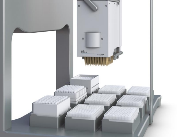
Agilent AssayMAP Bravo Liquid Handler
The AssayMAP Bravo Platform is a state-of-the-art Bravo liquid handler enhanced with a Bravo AM Head containing precision flow syringes. Liquid flow is precisely controlled to accommodate quantitative binding and elution in a single pass. The syringes are designed for use specifically with AssayMAP Sample Preparation cartridges which incorporate a 5 μL packed bed of resin supported by membranes molded into the polypropylene cartridge, enabling bidirectional flow and true high throughput chromatography. Recoveries approach 100% with reproducibility similar to HPLC (< 5% CV). A simple user interface allows assay developers to easily set key assay parameters without an automation engineer for error-free, push-button routine operation. With a broad range of chemistries and laboratory-tested protocols that can be flexibly combined, the AssayMAP technology simplifies the most challenging sample preparation workflows. Sample preparation cartridges are available for
- antibody purification,
- N-glycan sample preparation, and
- reverse phase peptide desalting
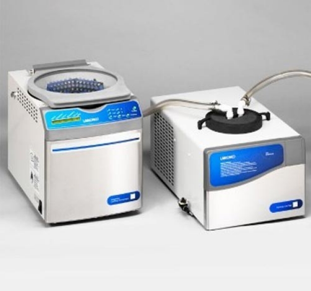
Labconco CentriVap Concentrator
The Labconco CentriVap Concentrator uses a combination of centrifugal force, vacuum and heat to speed evaporation of multiple small samples. As many as 148 samples may be processed at once. Sample sizes range from a few microliters up to 25 milliliters. The CentriVap is a component of a system that includes the CentriVap refrigerated cold trap and an acid-resistant vacuum pump. The cold trap helps protect the accessory vacuum pump from the corrosive effects of vapors and fumes as they evaporate from the samples.
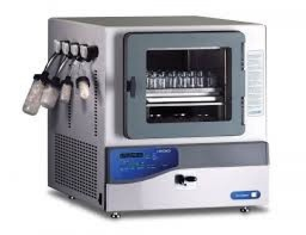
Labconco Triad Cascade Freeze Dry System
The Labconco Triad Cascade benchtop freeze dry system enables lyophilization either by tray drying with stoppering or conventional sample freeze drying with four sample valves on the left side. Samples for both types of freeze drying can be run at once. The chamber pre-freezes samples to save money and time, eliminating the need for a separate freezer and product transfer. User-friendly controls enable you to start runs quickly and the microprocessor controls up to five different ramping and holding segments to meet different temperature protocols. The hot gas defrost allows for easy cleanup of up to 1.8 liters of water or 2.5 liters of ice. The built-in pneumatic stoppering mechanism can handle glassware from 31mm to 148mm on its 12.4"w x 14.5"d shelf. Two 1/3 hp refrigeration systems cool the collector to -85° C (-121° F) to process low eutectic point samples including ones containing acetonitrile.
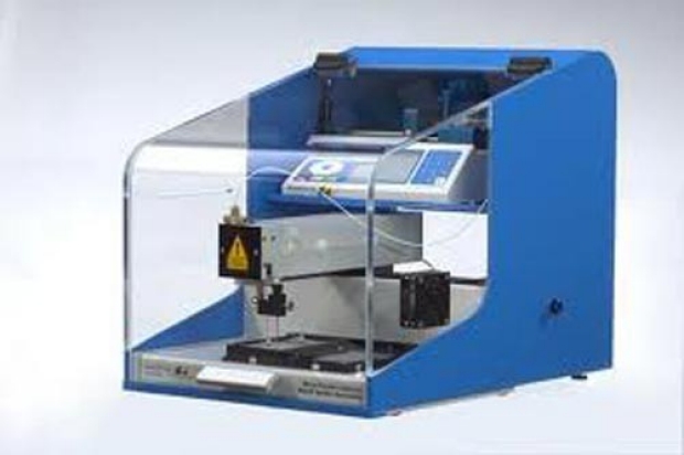
SunCollect MALDI Spotter-Sprayer
The SunCollect is a versatile instrument designed to operate as both an HPLC effluent touch-off spotter for MALDI target spotting and a micro-fraction collector for collection of HPLC effluent onto MALDI plates and into multiwall plates. For MALDI imaging mass spectrometry, The SunCollect offers a simple add-on accessory that allows for spraying protease and matrix solutions over the surface of a 2D tissue section by pneumatic atomization. Delivery of a homogeneous matrix layer is very reproducible and results in a very consistent matrix thickness. Depending on the matrix used, an initial light layer facilitates the formation of seed crystals that subsequently afford in a high degree of matrix homogeneity. System variables include matrix flow rate, needle speed, and number of layers. These can all be separately controlled over up to ten layers of matrix addition. The spraying software is simple and allows for quick changes to the speed of the spraying needle, number of tissue samples being sprayed, and the number of layers that will be placed over the tissue sections.
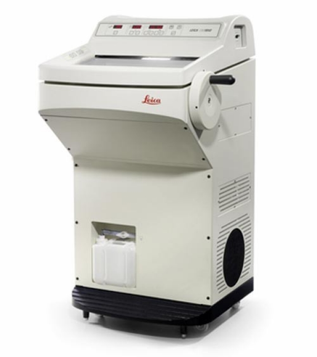
Leica CM1850 Cryostat
The Leica CM1850 cryostat design incorporates emphasis on power savings, increased efficiency and operator safety. The result is a versatile cryostat with an optimized cooling system, rapid specimen freezing and smooth specimen orientation for the high-quality sectioning demanded in routine histology and clinical pathology. The illuminated cryochamber is easily accessible through a large heated sliding window. Ample space is provided for convenient specimen handling. Smooth surfaces inside ensure safe and easy cleaning. The microtome is protected by a slot cover to allow easy and safe spray disinfection. The smooth specimen orientation with automatic centering and zero notch features outstanding handling capabilities for fast and precise sectioning.
A major challenge of mass spectrometry is data analysis. Capitalizing upon the Canary Center's bioinformatics resources, data generated by the proteomics core facility is archived and analyzed on a Dell Cluster housed in the core facility. This cluster provides 128TB of RAID storage, an 80 node computing cluster and web, database and file conversion servers. Users of the facility have convenient access to an easy-to-use, web-accessible, automated data analysis pipeline developed by Dr. Parag Mallick (based upon ProteoWizard and LabKey Server). Dr. Mallick is the principle investigator of the ProteoWizard projecta large software initiative that develops software for the analysis of mass spectrometry data. In 2013 alone, ProteoWizard was downloaded more than 42,000 times and is in use in labs across the world.
Proteomics Core Facility Description
The following description is suitable for use in conjunction with grant proposals:
The Proteomics Core Facility associated with the Canary Center at Stanford for Cancer Early Detection is a shared resource facility that supports Canary Center faculty, associates, and Canary Foundation science teams. The goal of the core facility is to enable a diverse group of investigators to leverage mass spectrometry in support of their research. This is accomplished through training and subsequent access to mass spectrometers at limited cost to Canary Center faculty, affiliates and their trainees. At present, the core facility supports four mass spectrometers including:
- Thermo Orbitrap Elite with ETD detector;
- Thermo LTQ Orbitrap Velos;
- Agilent 6490 triple quadrupole LC/MS; and
- AB SCIEX 5800 TOF/TOF (MALDI) systems.
Other instruments available in the MS Core include an Agilent AssayMAP Bravo liquid-handing robotic system and a Biacore X100 bimolecular interaction system. The core facility occupies over 2200 sq. ft. and the central room is designed to support ten LC-MS/MS systems including as many as six large LTQ Orbitrap-like systems. There are access panels in proximity to each mass spectrometer that provide nitrogen, UHP helium, compressed air, and an extractor for removal of vacuum pump fumes. Uninterrupted power supply (UPS) systems and an emergency backup generator support all mass spectrometers, HPLCs and computers. Adjacent to the central room is a sample preparation room (with balances, fume hood and water system) and a utility room (with flammable solvent storage and switching systems for nitrogen and UHP helium). An additional lab supports a MALDI mass spectrometer and instruments associated with MALDI applications including LC-MALDI and MALDI imaging mass spectrometry. A major challenge of mass spectrometry is the analysis of the data. Capitalizing upon the Canary Center's bioinformatics resources, data generated by the Proteomics Core Facility is archived and analyzed on a Dell Cluster housed within the Canary Center. This cluster provides 128TB of RAID storage, an 80 node computing cluster and web, database and file conversion servers. Users of the facility have convenient access to an easy-to-use, web-accessible, automated data analysis pipeline developed by Dr. Mallick (based upon ProteoWizard and LabKey Server).